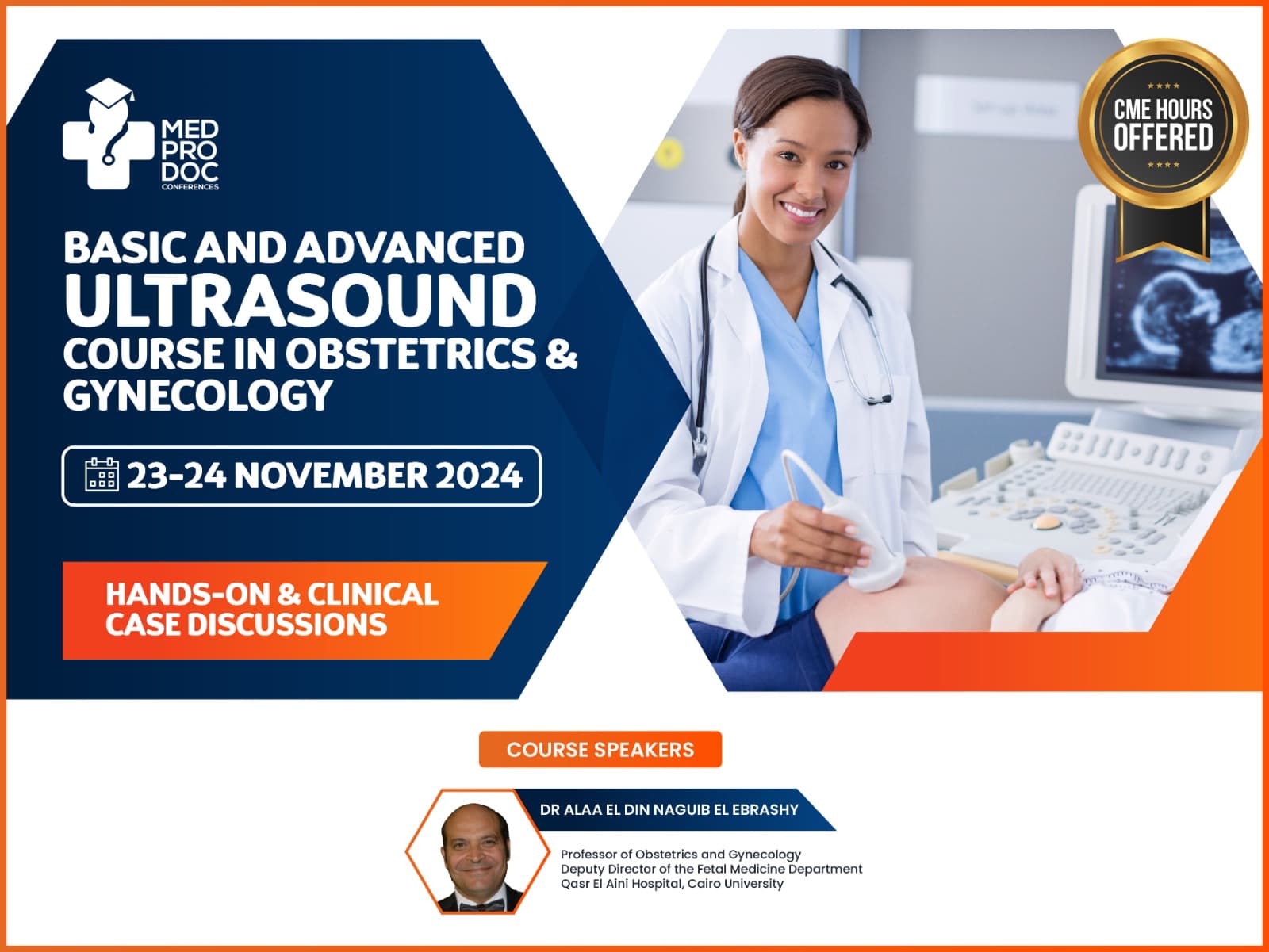
Basic and Advanced Ultrasound Course in Obstetrics & Gynecology
HANDS-ON & CLINICAL CASE DISCUSSIONS
Course Objective
Our upcoming 2024 Basic and Advanced Ultrasound Training Course in Dubai in Obstetrics and Gynecology is for doctors and other related medical professionals. Chiefly, it focuses on the provision of an all-inclusive understanding of Fetal Medicine & Ultrasound in Gynecology. It will be on November 23 – 24 organized by MED Prodoc Conferences Dubai, UAE, Approved by ISUOG, and Accredited by Dubai Health Authority(DHA).
The primary objective of this ultrasound training in Dubai is to equip healthcare professionals, particularly obstetricians and gynecologists, with comprehensive knowledge and practical skills in maternofetal medicine and gynecological ultrasound. By the end of the course, participants will be proficient in utilizing ultrasound techniques during pregnancy and gynecological examinations, adhering to international standards such as those outlined by the International Society of Ultrasound in Obstetrics and Gynecology (ISUOG).

COURSE DETAILS
Date : 31 May – 01 June 2025
Course Type: Hands-On & Clinical Case Discussion
Minimum Qualifications: MBBS
Who Can Attend: Obstetrics and gynecologist, sonographers, Ultrasound technicians & radiologists
HIGHLIGHTS OF THE ULTRASOUND COURSE IN DUBAI
-
Early Pregnancy Ultrasound:
- Conduct Sono embryology, therefore emphasizing complications like ectopic pregnancy and abortion.
- Focus on the 13-week scanning process with case demonstration in the ultrasound course, incorporating the latest applications of fetal anatomy checklists and screening for fetal aneuploidy and pre-eclampsia.
-
Mid-Trimester Ultrasound Case Demonstration:
- Teach the formulation of obstetric reports, including fetal biometry and growth assessment.
- Provide a comprehensive approach to fetal anatomy, emphasizing landmarks for diagnosing abnormal fetal morphology.
- In-depth discussion on placenta, liquor, and umbilical cord abnormalities, with a special focus on the morbid placenta.
-
Doppler Utilization:
- Understand Doppler techniques in our ultrasound course and accordingly integrate them into daily clinical practice in a simplified manner.
-
3D Ultrasound Applications:
- Demonstrate the application as well as benefits of 3D ultrasound for patient care.
-
Twin Pregnancy Ultrasound:
- Explore ultrasound perspectives on early, mid, and late follow-up of twins.
- Address specific conditions and challenges encountered in twin pregnancies.
-
Gynecological Ultrasound:
- Provide an in-depth understanding of uterine, ovarian, and adnexal ultrasound.
- Also familiarize participants with transvaginal ultrasound for uterine pathology, Mullerian abnormalities, ovarian cysts, and adnexal pathology.
- Early Pregnancy Ultrasound – Normal and Abnormal findings
Throughout the course, participants will have access to a wealth of materials, including papers and lectures, all aligned with ISUOG guidelines. Basically, the objective is to empower healthcare professionals with the knowledge and skills necessary to conduct effective maternofetal and gynecological ultrasound examinations, contributing to improved patient care and outcomes.
We provide hands-on practical training workshop and is undoubtedly among one of the best gynecologic ultrasound training course organization in Dubai, offering the world’s most modern state-of-art simulators with Accredited DHA Continuing Medical Education hours (CME) to upgrade knowledge and skills under internationally recognized certifications and faculties.
INTRODUCTION TO OBSTETRIC ULTRASOUND
-
How to Write an Obstetric Report
- Understanding the structure and key components
- Tips for clear and concise reporting
-
Fetal Central Nervous System (CNS) and Face
- Normal development and anatomy
- Identification of abnormalities in CNS and facial structures
-
Fetal Heart Examination
- Techniques for assessing fetal heart anatomy
- Differentiating between normal and abnormal finding
-
Fetal Gastrointestinal Tract (GIT), Renal, and Skeletal Systems
- Normal fetal GIT, renal, and skeletal development
- Recognition of abnormalities in these systems
ADVANCED TOPICS IN OBSTETRIC AND GYNECOLOGIC ULTRASOUND COURSE
-
13-Week Fetal Scan: Screening and Anatomy
- Importance of the 13-week scan in early pregnancy
- Detailed examination of fetal anatomy and screening protocols
-
Doppler in Obstetrics: Clinical Application
- Understanding Doppler ultrasound in obstetrics
- Clinical applications for assessing blood flow in various scenarios
-
Ultrasound for Twins: Practical Guidelines
- Techniques for imaging and assessing twins
- Challenges and considerations in twin pregnancies
-
Preterm Labor: Prediction and Management
- Identifying risk factors for preterm labor
- Strategies for prediction and effective management
-
How to Write a Gynecology Report
- Structure and content of gynecology reports
- Recognizing and reporting normal and abnormal findings in gynecologic ultrasound
ABOUT OBSTETRICS & GYNECOLOGY ULTRASOUND
The ultrasound image is also called ultrasound scanning or sonography. Obstetric ultrasound uses sound waves to obtain pictures of an embryo or fetus within a pregnant woman as well as the woman’s uterus and ovaries. This is a perfect method for monitoring a pregnant lady and their unborn babies, and the images are captured in real-time, so they can show the structure and movement of the body’s internal organs. They can also show blood flowing through blood vessels.
During an obstetric ultrasound, the examiner may evaluate blood flow in the umbilical cord or may, in some cases, assess blood flow in the fetus or placenta.
Ob-Gyn ultrasound imaging is a noninvasive medical test that helps physicians diagnose and treat medical conditions. Ultrasound is nondestructive testing of products and structures, also ultrasound is used to detect invisible flaws.
Our ultrasound training course lecture topic “first trimester of pregnancy” which is performed between 5th and 6th week able to see the gestational and yolk sacs and subsequently it allows your doctor to confirm your pregnancy. If your ultrasound is completed between weeks 6 and 7, the baby, which is now called an embryo is measuring nearly 9 mm in length and also a heartbeat is seen within this timeframe.
Ultrasounds scans during 8th to 11th weeks will show your growing baby, during this time the fetus has identifiable features such as the body, head, arms, and legs, and is moving quite readily within the gestational sac.
Med Prodoc ultrasound courses for doctors include guidelines for the second trimester and ultrasound procedures designed to assist practitioners in producing high-quality diagnostic surveys of the fetus by demonstrating and describing recommended audio-video imaging.


